2011年3月に日本で発生した3つの悲劇から10年経ち、原子放射線の影響に関する国連科学委員会(the United Nations Scientific Committee on the Effects of Atomic Radiation:UNSCEAR)は本日公表となる2020年報告書(2020Report)の中で、放射線被ばくが直接の原因となる健康影響(例えば発がん)が将来的に見られる可能性は低いと言及している。
表題“2011年東日本大震災後の福島第一原子力発電所における事故による放射線被ばくのレベルと影響:UNSCEAR2013年報告書刊行後に発表された知見の影響(Levels and effects of radiation exposure due to the accident at the Fukushima Daiichi Nuclear Power Station: Implications of information published since the UNSCEAR2013Report)”のUNSCEAR2020年報告書は、2019年末までに公表された関連する全ての科学的知見(査読付き論文と観測データ)をとりまとめている。これらは福島第一原子力発電所(福島第一原発)の事故による放射線被ばくのレベルと影響に関連するものである。本報告書の目的は全科学的知見をとりまとめ、UNSCEAR2013年報告書についてこれら知見の影響を評価することである。全体的にみると、2020年報告書はUNSCEAR2013年報告書の主な知見と結論を概して確認するものであった。
Numerical Simulation Based on Individual Voxel Phantoms for a Sophisticated Evaluation of Internal Doses Mainly From 131I in Highly Exposed Workers Involved in the TEPCO Fukushima Daiichi NPP Accident. 2019 Health Phys.116:647-656
An accident of internal contamination with plutonium and americium at a nuclear facility in japan: a preliminary report and the possibility of dtpa administration adding to the diagnosis. 2018 Radiat Prot Dosimetry182:98-103
Experiences of population monitoring using whole-body counters in response to the Fukushima nuclear accident. 2018 Health Phys 115:259-274
Early Intake of Radiocesium by Residents Living Near the Tepco Fukushima Dai-ichi Nuclear Power Plant After the Accident. Part 2: Relationship Between Internal Dose and Evacuation Behavior in Individuals. 2017 Health Phys 112:512-525
The first meeting of the who guideline development group for the revision of the WHO 1999 guidelines for iodine thyroid blocking 2016 Radiat Prot Dosimetry 171:47-56
2020年 8月 IAEA Webinar Material – WEBINAR on medical response to nuclear and radiological safety or security related emergencies: Lessons learned from case studies
2019年 5月 IAEA Regional Workshop on Medical Preparedness and Response in case of a Nuclear or Radiological Emergency(ウルグアイ)
2019年 2月 IAEA UAE National Training Course on Medical Preparedness and Response to Radiation Emergencies in coordination with the IAEA and NCEMA(アラブ首長国連邦)
2018年11月 IAEA Expert Mission to Review Medical Preparedness to Radiological Emergency(アラブ首長国連邦)
2018年 5月 WHO The 3rd Asian WHO/REMPAN Workshop/Monitoring, Assessment and Management of Internal Contamination(韓国)
Mainly on the Chromosome Aberrations Induced by Tritium
Tada-aka HORI*1 and Sayaka NAKAI*1
*1放射線医学総合研究所遺伝研究部;千葉市穴川4-9-1
(〒280)
Division of Gerletics, National Institute of Radiological
Sciences; 9-1, Anagawa 4℃home, Chibashi,
Chiba-ken.
Genetic risk assessment for potential hazard from environmental tritium to man becomes important with increasing nuclear-power industry. The purpose of this short review is to discuss the possible genetic effects of tritium from a view of genetic risk estimation.
The discussion is based mainly on our experimental results on the chromosome aberrations induced in human lymphocytes by tritium at the very low-level. The types of chromosome aberrations induced by radiation from tritium incorporated into the cells are mostly chromatid types. The most interesting finding is that the dose-response relationship observed in both tritiated-water and tritiatedthymidine is composed of two phases. The examination on the nature of two-phase dose-response relationship is very important not only for the mechanisms of chromosome aberrations, but also for the evaluation of genetic risk from low-level radiation.
1) J. J. COHEN and G. H. HIGGINS; The socioeconomic impact of low-level tritium releases to the environment, “Tritium, ” ed. A. A.MOGHISSI and MW. CARTER, 14 (1973).
2) “Tritium, ” ed. A. A. MOGHISSI and MW.CARTER (1973).
3) United Nations, Report of the United Nations Scientific Committee on the Effects of Atomic Radiation, Ionizing Radiation: “Levels and Effects, ” Vol. II, Effect, New York (1972).
4) BEIR Report, The Effects on Populations of Exposure to Low Levels of Ionizing Radiation.
Report of the Advisory Committee on the Biological Effects of Ionizing Radiation. National Academy of Sciences, National Research Council, Washington, D. C., (1972).
5) K. SAX; An analysis of X-ray induced chromosome aberrations in, Tradescantia, Genetics,25, 41 (1940).
6) D. E. LEA; Actions of Radiation on Living Cells, 2nd ed., Cambridge University Press(1955).
7) H. J. EVANS et al.; Human Radiation Cytogenetics,North-Holland Publishing Co. (1967).
S) A. L. BROOKS et al.; Effect of 239PuO2 particle number and size on the frequency and distribution
of chromosome aberrations in the liver of the Chinese hamster, Radiat. Res.,59, 693 (1974).
10) FT. HATCH and J. A. MAZRIMAS; Tritiation of animals from tritiated water, Radiat. Res.,50, 339 (1972).
11) A. M. UENO; Incorporation of tritium from tritiated water into nucleic acids of Oryzias latipes eggs, Radiat. Res., 59, 629 (1974).
12) J. E. CLEAVER et al.; Biological damages from intranuclear tritium: DNA strand breaks and their repair, Science, 177, 996 (1972).
13) J. TOMIZAWA and T. OGAWA; Breakage of polynucleotide strands by disintegration of radioposphorous atoms in DNA molecules and their repair. II. Simultaneous breakage of both etrarde, J. Mol. Biol., 30, 7 (1967).
14) J. H. TAYLOR; Sister chromatid exchanges in tritium-labeled chromo omen, Genetics, 43, 515 (1958).
15) D. E. WIMBER; Chromosome breakage produced by tritium-labeled thymidine in Trades cantia paldosa, Proc. Natl. Acad. Sci. U. S. A.,45, 839 (1959).
16) H. A. MCQUADE and M. FRIEDKIN; Radiation effects of thymidine 3H and thymidine ‘4C, Exptl. Cell Res., 21, 118 (1960).
17) A. T. NATARAJAN; Chromosome breakage and mitotic inhibition induced by tritiated thymidine in root meristems of Vicia faba, Exptl.Cell Res., 22, 275 (1961).
18) M. A. BENDER et al.; Aberrations induced in human lymphocyte chromosomes by 3H-labeled nucleotides, Cytogenetics, 1, 65 (1962).
19) K. V. BALDEV; Lack of relationship between chromosome aberrations induced by and localized incorporation of 3H-TdR in human leucocytes, Internatl. Congr. Radiat. Res., 206 (1974).
20) CM. OCKEY; Chromatid aberrations resulting from 3H-thymidine incorporation into early and late S periods in human fibroblasts, Int. J. Radiat. Biol., 13, 479 (1967).
21) R. R. KLEVECZ and T. C. Hsu; The differential capacity for RNA synthesis among chromosomes:
a cytological approach, Proc. Natl. Acad. Sci. U. S. A., 52, 811 (1964).
22) J. G. BREWEN and G. OLIVIERI; The kinetics of chromatid aberrations induced in Chinese hamster cells by tritium-labeled thymidine, Radiat. Res., 28, 779 (1966).
23) W. C. DEWEY et al. : Comparisons of tritiated thymidine, tritiated water, and cobalt-60 gamma rays in inducing chromosomal aberrations, Radiat. Res., 24, 214 (1965).
24) C. R. GEARD; Chromosomal aberrations in three successive cell cycles of Wallabia bicolor leucocytes after tritiated thymidine incorporation, Radiat. Res., 61, 118 (1975).
25) T. KUROIWA and N. TANAKA; Effect of THOwater on Crepis chromosomes, Jap. J. Genet., 47, 356 (1972).
26) S. PERSON and R. C. BOCKRATH, Jr.; Differential mutation production by the decay of incorporated tritiated compound in E. coli, Biophys. J., 4, 355 (1964).
27) W. D. KAPKAN and J. E. SISKEN; Genetic and autoradiographic studies of tritiated thymidine in testes of Drosophila melanogaster, Experimentia, 16, 67 (1960).
28) O. STROMNAES; Mutation effect of C14 and H3 labeled DNA precursors injected into Droso phila melanogaster males, Canad. J. Cytol. Genet., 4, 440 (1962).
29) W. D. KAPLAN et al.; Nonrandom distribution of lethals induced by tritiated thymidine in Drosophila melanogaster, Genetics, 49, 701 (1964).
30) P. KIEFT; Induction of recessive lethals by 3H-uridine and 3H-thymidine in Drosophila. Biological Effects of Transmutation and Decay of Incorporated Radioisotopes (Proc. Panel, Vienna, 1967), IAEA, Vienna, 65 (1968).
31) O. STROMNAES and I. KVELLAND; The induction of minute mutations in Drosophila with tritium-labeled thymidine, Genetics, 48, 1559 (1963).
32) A. J. BATEMAN and A. C. CHANDLEY; Mutations induced in the mouse with tritiated thymidine, Nature, 193, 705 (1962).
33) R. B. CUMMING et al.; Tritium-induced specific locus mutation rate in cells of the male mouse, Internatl. Congr. Radiat. Res., 63 (1974).
34) United Nations, Report of the United Nations Scientific Committee on the Effects of Atomic Radiation. General Assembly Official Records: Genetic Effects of Radiation (1975).
35) P. PERRY and H. J. EVANS; Cytological detection of mutagen-carcinogen exposure by sister chromatid exchange, Nature, 258, 121 (1975).
36) T. HORI and S. NAKAI; Chromosome aberrations induced in human lymphocytes by tritiated water at low level, J. Radiat. Res., 16, 57 (1975).
UNUSUAL DOSE–RESPONSE OF CHROMOSOME ABERRATIONS INDUCED IN HUMAN LYMPHOCYTES BY VERY LOW DOSE EXPOSURES TO TRITIUM TADA-AKI HORI and SAYAKA NAKAI 1978年
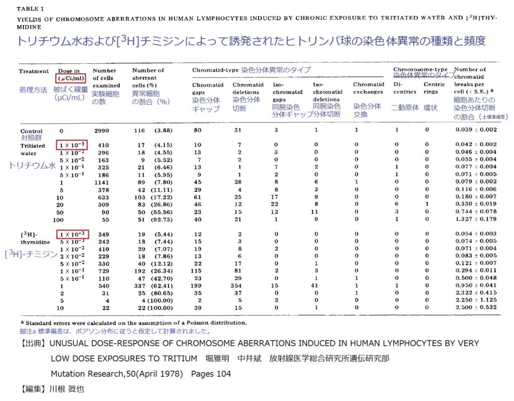
細胞あたりの染色分体切断の割合を計算したものが、実験対象と方法(Materials and methods)表1の最後の欄に記載されています。トリチウム(μCi/mL)の被ばく線量と細胞あたりの染色分体切断数(対照群の同切断数を差し引いています)との関係は図2に示されています。線量に対する切断数は対数目盛で示されています。[3H]チミジンは、トリチウム化水よりおよそ100倍染色体異常を生じる効果的があるように見えます。被ばく線量は細胞培養の際のトリチウム濃度を意味するので、[3H]チミジンの一見の高い線量効果はトリチウムが染色体DNAに取り込まれることによる非常に局所的なベータ線照射によって引き起こされた、と解釈することができます。
その線量―応答関係は両方のケースで変わった曲線を描きました。これは注目に値します。染色体異常が生まれる率はトリチウム水の場合は5μCi/mL以下で、[3H]チミジンの場合は5×10-2μCi/mL以下でそれぞれより少なくなっています。我々はこの2つの曲線について、べき乗数則(the power low model)Y = k D nに基づいて、最小二乗法回帰分析を行いました。Yが細胞あたりの染色分体切断数であり、DはμCi/mLで表された被ばく線量、kとnは定数です。
対数変換すると、この関係は直線、log Y =log k+ n log D となります。表2で示すように、べき乗数則は、データに極めて良く適合しています。統計分析のこれらの結果から、我々は、48時間のトリチウムへの慢性暴露によって誘発される染色体異常の線量―応答関係は、2つの要素を持っているととりあえず結論しました。高い線量での線量―応答関係では、線量D のべき指数nは、トリチウム水では0.953と[3H]チミジンでは0.790と1.0から大きく外れず、線量に対する細胞あたりの切断数が線量に直線的に依存することを示しています。P値については、トリチウム水では0.1>P> 0.05、[3H]チミジンでは0.3>P> 0.2でした。しかし、低い線量の範囲では、線量D のべき指数nがトリチウム水では0.380、[3H]チミジン0.338であり1.0からかなり逸脱して、明らかに線形動力学に当てはまりません。このときP値はP < 0.001でした。したがって、部分的ヒットまたは部分的ターゲットによって引き起こされることが明らかになりました。トリチウム水と[3H]チミジンにいずれにおいても、細胞の染色分体切断の数は、いずれの線量においてもポアソン分布を示しました。ただし[3H]チミジンの50μCi/mLおよび10μCi/mLの2つのケースは例外です。ここでは選択的な細胞死が恐らく起こるかもしれません。したがって、極めて低い線量の範囲で示された変わった線量依存関係は、染色体異常の偏った記録とする根拠にはならないかもしれません。
地球上に生きる動植物の生命を維持する上でなくてはならないものの一つである水は,人間の体重の60~70%を占めている.水には水素の放射性同位体であるトリチウムが含まれており,このトリチウムは半減期12.3 年でβ壊変してヘリウム3になる放射性核種である.トリチウムは大気上層において,宇宙線(陽子や中性子)と大気を構成する窒素原子や酸素原子との核反応により,定常的に生成されており,その量は年間200 g 程度と見積もられている[1].大気中で生成した天然トリチウムのほとんどは速やかに酸化されて水になり,やがて対流圏に移動して雨として地表面に降下する.トリチウムは空気中の水蒸気,雨,海水や地表水などに広く存在して水と一緒に自然界を循環しているため,大昔から人は環境中のトリチウムを飲料水あるいは食物として摂取してきた.光合成を出発点とするトリチウムの有機物への変換は,トリチウムの環境サイクルの重要な部分を占め,食物連鎖を介して人へトリチウムが移行する.
被ばく線量評価に関連するトリチウムの環境サイクルを明らかにするためには,植物等の有機物中に存在するトリチウム濃度測定が不可欠である.有機物中に存在するトリチウムは組織自由水トリチウム(Free Water Tritium:FWT)とOBT がある.これらは図3に示すように,凍結乾燥等により組織自由水と乾燥試料に分けて分析を行う.FWT濃度を分析する際,組織自由水を回収し,過マンガン酸カリウム,過酸化ナトリウムを加えて蒸留し,その中に含まれる有機物を分解除去して精製する必要がある.分
や海草,魚のFWT,OBT 濃度の増加が認められている[22,26].また,気体廃棄物として排気筒から環境へ放出されたトリチウムの一部は,施設近傍において直接沈着や雨で降下する.例えばフランスのValdacにある施設周辺では環境トリチウム濃度レベルが上昇し,その結果樹木や地衣類(lichen)のOBT 濃度が上昇していることが確認されている[27,28].日本では茨城県東海村の核燃料再処理施設周辺で大気,葉菜のモニタリング例があり,施設から半径5 km 以内にトリチウム濃度の上昇が認められている[29].
5.4 核融合実験施設
核融合実験施設の一例として,イギリスのカラム研究所にあるJET(Joint European Torus)やアメリカのプリンストン大学にあったTFTR(Tokamak Fusion Test Reactor)が挙げられる.これらの実験施設では重水素とトリチウムを用いた核融合プラズマ実験が行われた.その間,施設周辺における大気HTO 濃度をパッシブサンプラーを用いて評価を行った例[30,31]や,降水や陸水のモニタリングを行った報告がある[32].いずれも施設近傍においてバックグラウンドに対して有意に高いレベルを示していた.
[1]UNSCEAR, Sources and Effects of Ionizing Radiation, vol.1 (United Nations Publications, New York, 2000).
[2]http://www.kankyo-hoshano.go.jp/data.html.
[3]http://www.nirs.go.jp/db/anzendb/NetsDB.html#.
[4]ICRP, Age−dependent Doses to Members of the Public from Intake of Radionuclides : Part5 Compilation of Ingestion and Inhalation Dose Coefficients, vol. 72 (Pergamon Press, Oxford,1995).
[5]N. Momoshima et al., Fusion Sci. Technol. 48, 520 (2005).
[6]N. Akata et al., Fusion Sci. Technol. 60, 1292 (2011).
[7]T. Iida et al., Radiat. Prot. Dosim. 58, 23 (1995).
[8]M. J. Wood, Health Phys. 70, 258 (1996).
[9]M. J. Wood and W. J. G. Workman, Fusion Technol. 21, 529(1992).
[10]N. Akata et al., J. Environ. Radioact. 102, 837 (2011).
[11]T. Okai et al., J. Radioanal. Nucl. Chem. 239, 527 (1999).
[12]T. Tamari et al., Fusion Sci. Technol. 60, 1252 (2011).
[13]S. Diabate and S. Strack, Health Phys 65, 698 (1993).
[14]H. Kakiuchi et al., Fusion Sci. Technol. 60, 1256 (2011).
[15]S. Ueda et al., J. Radioanal. Nucl. Chem. 267, 29 (2005).
[16]K. Shinotsuka et al., J. Radioanal. Nucl. Chem. 258, 233(2003).
[17]N. Momoshima et al., J. Nucl. Radiochem. Sci. 8, 117 (2007).
[18]F. Luykx and G. Fraser, Radiat. Prot. Dosim. 16, 31 (1986).
[19]T. Kotzer and A. Trivedi, Radiat. Prot. Dosim. 93, 61 (2001).
[20]T. G. Kotzer and W. J. G. Workman, Measurements of Tritium(HTO, TFWT, OBT) in Environmental Samples at Varying Distances from a Nuclear Generating Station, Atomic Energy of Canada Limited Report, AECL-12029 (Chalk River Laboratories, Chalk River, Canada, 1999).
[21]J. T. Harris et al., Health Phys. 95, 203 (2008).
[22]M. Masson et al., Radioprotection 40, S621 (2005).
[23]A. Baeza et al., J. Environ. Radioact. 86, 367 (2006).
[24]A. Baeza et al., J. Environ. Radioact. 100, 209 (2009).
[25]H. Hayakawa et al., “Proc. IRPA-10, Hiroshima, P-4a-235(2000),
http://www.2000.irpa.net/irpa10/cdrom/00608.pdf.”
[26]MAFF and SEPA, Radioactivity in Food and the Environment,1997 (RIFE−3) (Ministry of Agriculture, Fisheries and Food, and Scottish Environment Protection Agency,London, 1998).
[27]O. Daillant et al., Sci. Total Environ. 323, 253 (2004).
[28]L. Vichot et al., J. Environ. Radioact. 99, 1636 (2008).
[29]H. Fujita et al., J. Nucl. Sci. Technol. 44, 1474 (2007).
[30]R. L. Otlet et al., Fusion Technol. 21, 550 (1992).
[31]B. Patel et al., Fusion Eng. Des. 47, 267 (1999).
[32]V. L. Finley, PPPL-4481, (Princeton Plasma Physics Laboratory,

 セシウム137-1.9 2021年6月10日-14:00 田中幸博-2-743x1024.jpg)
 セシウム137-1.9 スペクトルデータ 2021年6月10日-14:00 田中幸博-804x1024.jpg)


」 榊原崇仁 pp237-1-555x1024.jpg)
と3Hーチミジン(3HーTdR)によって誘発された染色体異常の種類とその頻度 堀雅明 中井斌 1976年-1024x627.jpg)
濃度 1960~2011年.jpg)
